Videonystagmography (VNG): A Diagnostic Window into Brainstem and Vestibular Function
Videonystagmography (VNG) is a non-invasive diagnostic tool used to evaluate involuntary eye movements (nystagmus) in response to visual and vestibular stimuli. These eye movements are tightly linked to the function of the brainstem, cerebellum, and inner ear (vestibular system), making VNG a critical tool for assessing neurological integrity.
How VNG Works
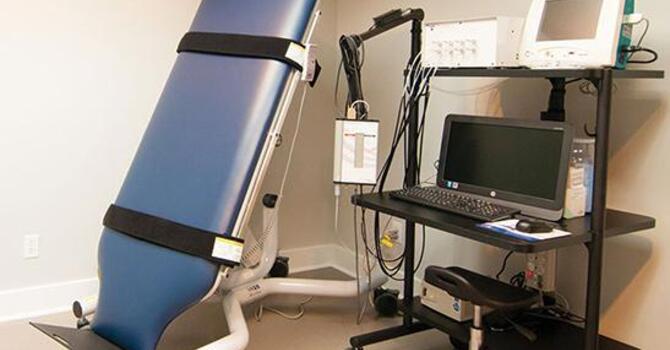
During a VNG test, a patient wears infrared goggles that track and record eye movements in various conditions:
-
Reflexive and Volitional Oculomotor Testing
-
Evaluates subconscious eye movement activity, gaze fixation (being able to hold your eyes still) in the dark, and optokinetic or rotational responses all being in the dark and without a target or object to look at.
-
These movements reflect the integrity of the brainstem and cerebellar circuits involved in eye movement control.
-
-
Positional Testing
-
Assesses eye movements when the head or body is placed in specific positions.
-
Detects positional nystagmus often linked to benign paroxysmal positional vertigo (BPPV) or central vestibular disorders.
-
What VNG Reveals About Neurological Function
-
Brainstem Integrity:
Caloric and oculomotor tests evaluate reflexes mediated through the brainstem, particularly the vestibular nuclei, medial longitudinal fasciculus, and cranial nerve pathways. Abnormal responses can indicate dysfunction or injury in these regions. -
Cerebellar Function:
Abnormalities in smooth pursuit or gaze-holding suggest dysfunction in the cerebellar flocculus and vermis—key areas in stabilizing eye movements. -
Vestibular System Function:
VNG helps differentiate between peripheral vestibular disorders (e.g., vestibular neuritis, BPPV) and central causes (e.g., brainstem stroke, concussion-related dysregulation). -
Reflexive Control & Sensorimotor Integration:
Eye movement patterns provide insight into how the brain integrates sensory input and executes reflexive motor output—essential in assessing neuroplasticity and recovery potential.
How Functional Neurologists Use VNG
For a functional neurologist, VNG is a critical component of a comprehensive neurological exam. It allows for:
-
Precise Localization of Dysfunction:
VNG can distinguish between peripheral (inner ear) and central (brainstem/cerebellum) lesions, helping guide targeted rehabilitation strategies. -
Objective Measurement of Progress:
Repeated VNG testing can monitor changes over time, allowing practitioners to adjust treatment protocols based on quantifiable neurophysiological data. -
Customized Rehabilitation Plans:
Based on VNG findings, a functional neurologist may prescribe specific oculomotor, vestibular, or cerebellar rehabilitation exercises aimed at re-establishing normal brainstem and reflexive function. -
Post-Concussion Syndrome & Dysautonomia Management:
In patients with head injuries, abnormal VNG results often correlate with dysautonomia, dizziness, and visual instability—common symptoms that can be improved with targeted therapy.

Forrest Fisher
Contact Me